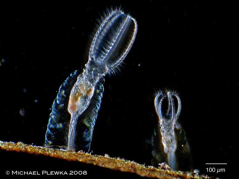
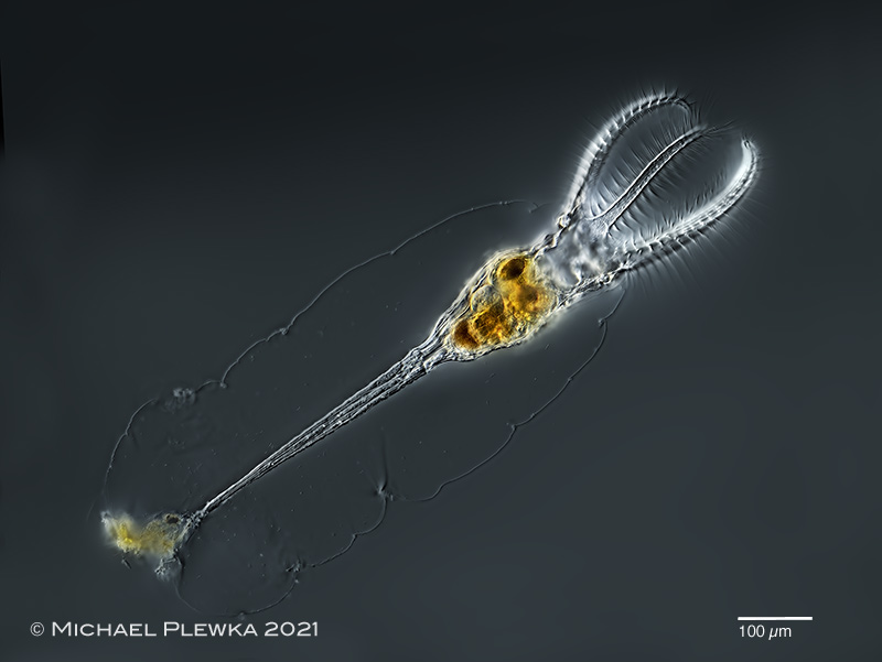
| Stephanceros fimbriatus, a sessile rotifer with a conspicuos corona and a gelatious sheath that is produced in periodic intervals by glands at the anterior / upper part of the foot. (1) |
| |
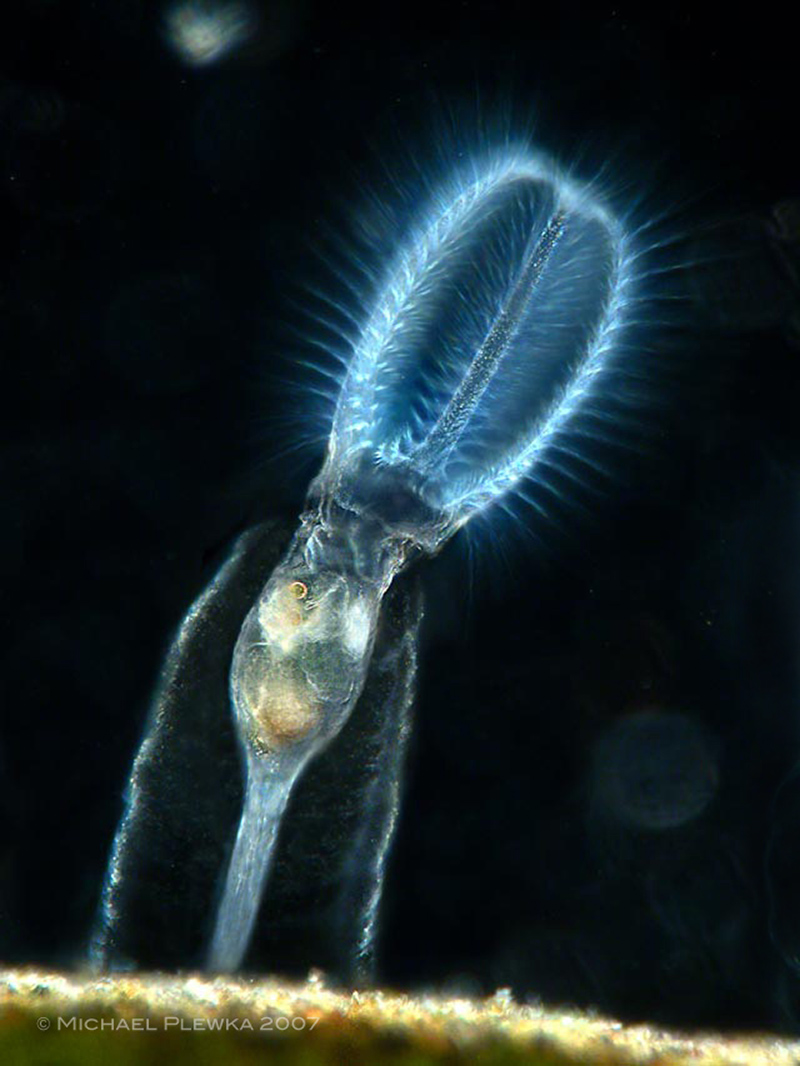 |
| Stephanceros fimbriatus, same specimen from above image. This rotifer lives in a mucilagineous sheath that is secreted by the integument. (1) |
| |
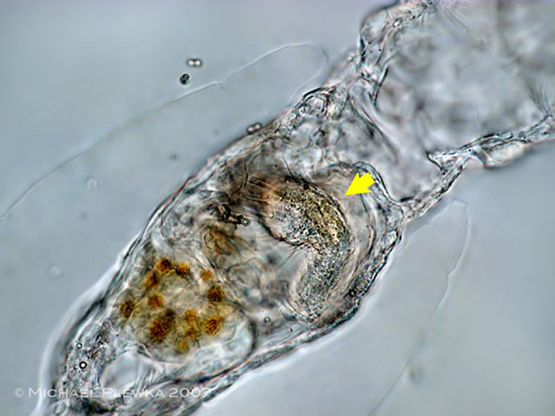 |
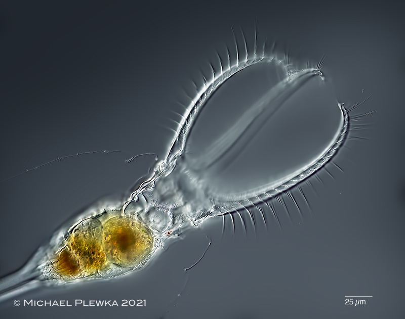
| Stephanceros fimbriatus; the food spectrum comprises small metazoans, for example hairybacks. In this image a hairyback (Gastrotrich) of the genus Chaetonotus is captured in the mastax (arrow). (1) |
| |
 |
| Stephanceros fimbriatus; specimen from (2) |
| |
 |
| Stephanceros fimbriatus; same specimen, focus plane on the proventriculus and stomach. The ventral side of the specimen is on the top side of the image.(2) |
| |
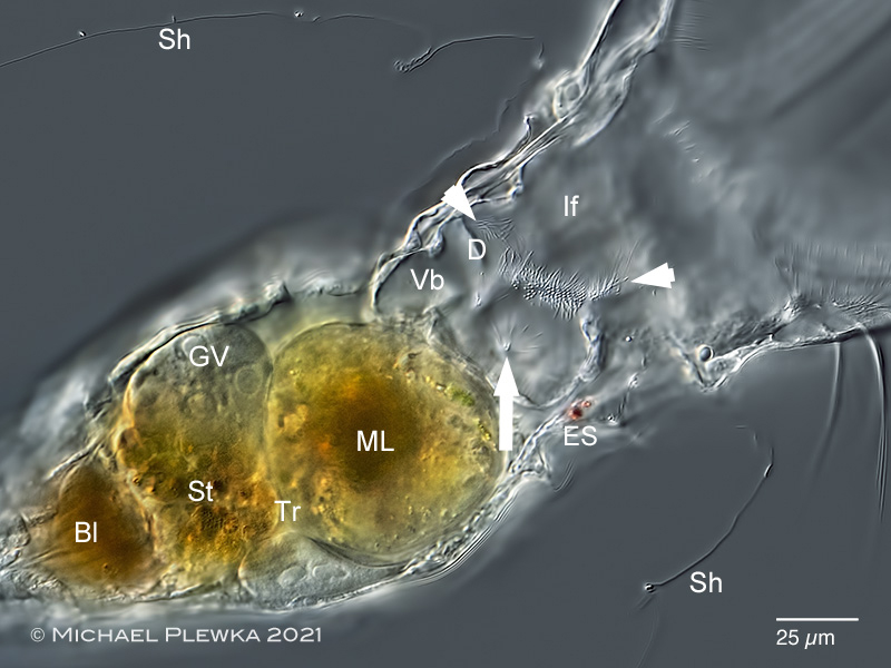 |
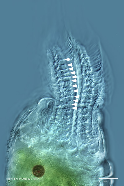
| Stephanceros fimbriatus; crop of the above image. The arrowheads mark a row of cilia (trochus after Donner (1965)) which are in permanent movement when the infundibulum is open. The arrow points to a group of (??sensory??) cilia that do not move when the infundibulum is open. Bl. bladder; D. diaphragma; ES: eyespot; GV: gemovitellarium; If: infundibulum; ML: mastax lumen; Sh: mucilaginous sheath; St. stomach; Tr: (location of the ) trophi (not visible); Vb: vestibulum; (2) |
| |
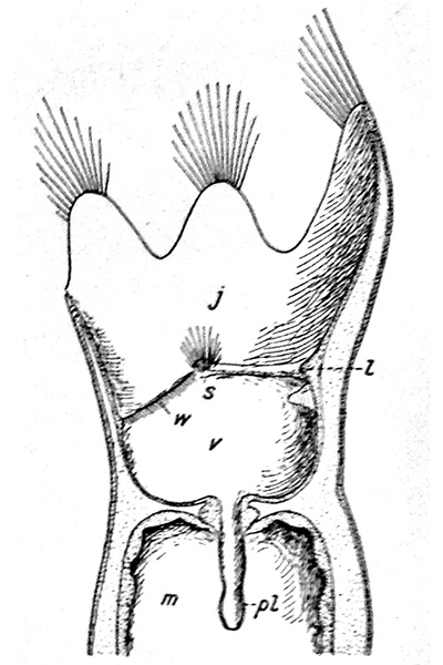 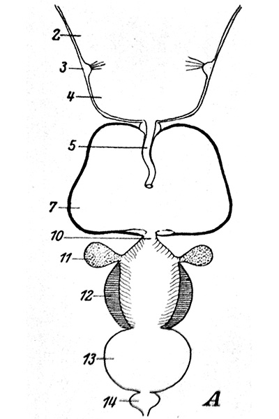 |
Head and digestive tract of collothecid rotifers; left image: Collotheca sp. ; right image: Stephanoceros fimbriatus; both images from Remane (1929). In principle, the morphology and function of the organs of the head region and the digestive system should be identical and thus homologous in the genera Collotheca and Stephanoceros. Now the caption to these pictures is interesting:
left picture (excerpt) : "...j: Infundibulum; l: Diaphragma; m: Pharynx oder Mastax; s: Sinnesorgan des Schlundrohrs, (sog. Pharyngealsinnesorgan); v: vestibulum; w: ventraler an das Diaphragma anschließender Wimpersaum."
right image (excerpt): "....2 Vestibulum; 3 Pharyngealsinnerorgan; 4 Infundibulum; 5: Pharyngealrohr; 7:Pharynx oder Mastax..."
So in the left picture, the infundibulum is the 1st section of the digestive tract, followed by the vestibulum, which opens into the mastax lumen via the pharyngeal tube. In the right picture it is the other way round: the vestibulum is the 1st section, the infundibulum is the 2nd section.
Thus there is a contradiction in the designation of the organs.
Both illustrations served as models for later authors; for example, in the book of Koste (1978) there is an illustration of Collotheca trilobata which takes the designation from the left picture; in a rotifer chapter of of DeSmet / Fontaneto (2015) the designation of the right picture is taken over.
This is all the more regrettable as in other works by Hochberg et al. (2018) l the entire "head" area is referred to as the infundibulum, for whatever reason. |
| |
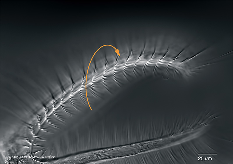 |
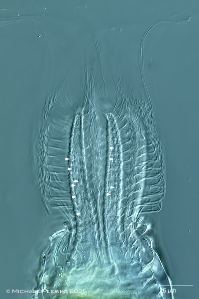
| Stephanceros fimbriatus; the bristles are arranged in the manner of a spiral staircase. If one of the bristles is stimulated a metachronous movement of the next bristles can be observed in the direction depicted by the arrow. |
| |
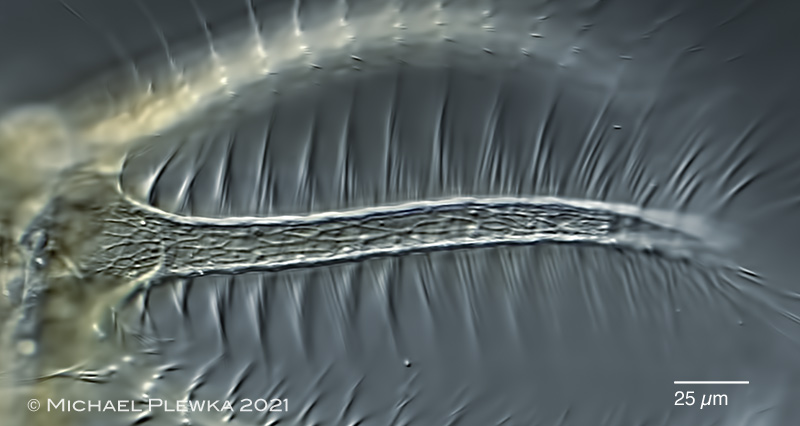 |
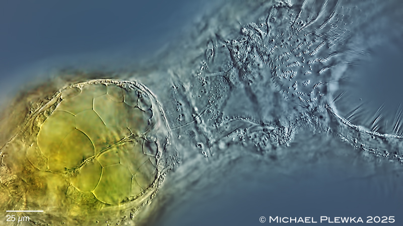
| Stephanceros fimbriatus; one of the 5 tentacles, focal plane on the net-like muscle fibers. |
| |
  |
| Stephanceros fimbriatus; two images of the retracted tentacles (excerpt from video). left image: when the tentacles are retracted into the trunk a segmentation of the tentacles becomes visible (segments marked by arrowheads) . Maybe these segments are functional units of the bristle movement. Right image: when the retractor muscles are released the tentacles protrude again while the bristles are moving, the outer bristles forming a rectangular shape. Focal plane on some ellipsoid bodies in the tentacles (arrowheads). |
| |
 |
 |
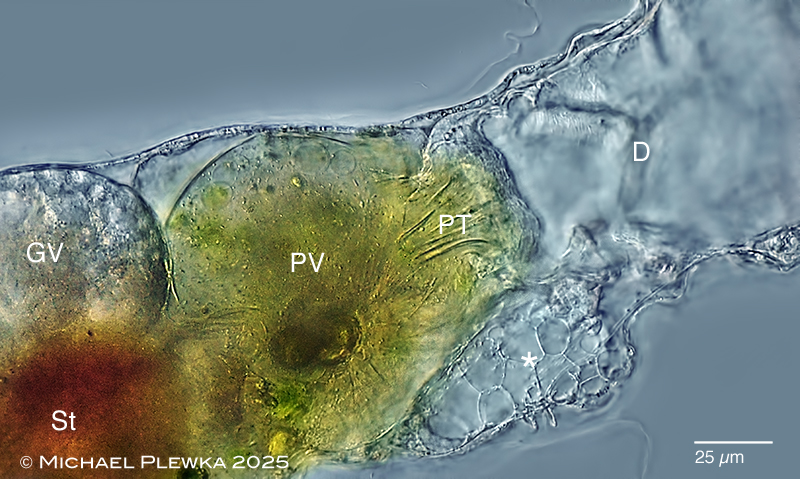
Stephanceros fimbriatus; two images of a structure of unknown function. Upper image: lateral view; the asterisk marks this structure which is on the dorsal side of the trunk. (D. diaphragma; GV: germovitellarium; PV: proventriculus / mastax lumen; PT: pharyngeal tube; St: stomach.
Lower image: dorsal view; on the left side the "large clear cells" of this organ are visible. Leydig (1855) calls them "eigenthümliches Organ); Hudson et Gosse (1886) believed that this organ is a nervous ganglion. De Beauchamp (1909) describes this structure as retrocerebral sac. Wesenberg-Lund (1929) calles it retrocerebral organ (which, by todays standards, is something different: a combination of two glands: retrocerebral sac and subcerebral glands.. |
| |
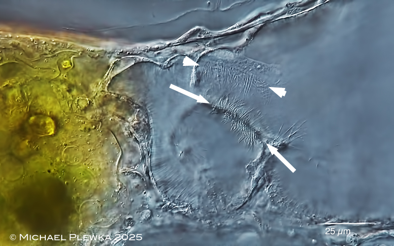 |
| Stephanceros fimbriatus; another optical transect of the diaphragma and its cilia in lateral view. A highspeed video (300 fps, still image here from this footage) reveals that the cilia marked by arrows are in dexioplectic metachronal movement which suggests that this structore might be homologous to the trochal cilia of the Philodinida / Gnesiotrocha. Also the structure marked by arrowheads shows diaplectic metachronal movement, whereas the other cilia at the outer walls of the vestibulum / infundibulum move only very unconspicuously. Dexioplectic metachronal movement means that the effective stroke of the cilia is directed to the right in this image which means that there is a current to the outside which is confirmed by the movement of small particles being driven out of the vestibulum. Like in Collotheca species the function of this current might be the transport of chemical attractants to the outside to attract potential prey like eugleind flagellates for example. |
| |
| |
| Collection of sample (2) courtesy of Uli Drabiniok |
| |
| |
|
|
| |
| Location: Wiedenbrück, pond (1); Nature reserve NSG Heiliges Meer, NRW, Germany, pond: Kleinweiher "Üffings Wiese" (location 16) (2); |
| Habitat: Epibiontic on Ceratophyllum sp. (1); plankton with detritus/ algae, together with Notommata cerberus and Lecane mira (2) |
| Date: 06.06.2008 (1); 16.11.2021 (2) |
|